
Microspore embryoGENESIS: A path to life or death

In vitro plant regeneration holds great potential in the agriculture and horticulture, especially in crop improvement and hybrid breeding. Moreover, it is also applicable to studies of plant developmental regulatory mechanisms. For in vitro embryogenesis, the most important step is to re-establish the totipotency and further develop into an embryo. Embryo cell fate can be established from different tissue culture. Among the different pathways, we focus on microspore embryogenesis, where haploid embryos arise from stress-induced microspore culture.
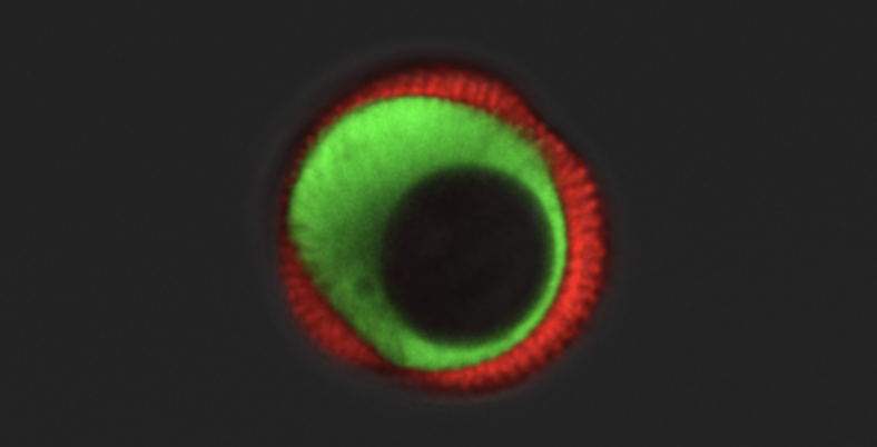
Microspores are pollen-precursor cells in the anther (Fig. 1), which are developmentally programmed to form gametes. However, under certain stress conditions, the isolated microspores can be re-programmed to undergo appropriate cell divisions and develop into an haploid embryo, even in the absence of fertilisation (Fig. 2).
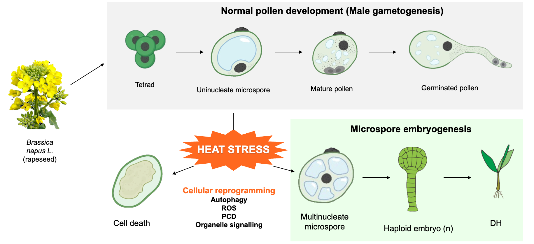
We use rapeseed (Brassica napus L.) as a model species to study microspore embryogenesis, where heat shock causes the microspores to change its developmental fate from pollen development to either developmental arrest, cell death, or embryogenesis (Corral-Martinez et al., 2020). Our lab focuses on exploring the role of autophagy during microspore embryogenesis, as puncta of ATG5 and ATG8 were observed in stress-induced microspore cultures in Horedum vulgare (barley) and Brassica napus (Pérez-Pérez et al., 2019). Autophagy is a self-eating pathway, which is the main route for degradation and recycling of cellular compartments. Therefore, autophagy might play an important role in cellular reprogramming of plant somatic cells from one developmental pathway to another (Rodriguez et al., 2020)
Team GENESIS consists of experts in plant regeneration and hormone signalling (Geelen lab) and 4 labs from Plant Systems Biology: Advanced Live Cell Imaging (Vandamme lab), Programmed Cell Death (Nowack lab), Oxidative Stress Signaling (Van Breusegem lab), and Inter-organelle Signalling (De Clercq lab). Together, we provide a concerted molecular approach to understand the mechanism in a cellular level behind microspore embryogenesis. The outcome of the project will lead to a solid molecular framework of androgenesis and novel, effective strategies for boosting the efficiency of stress-induced microspore embryogenesis and its wider application in hybrid breeding.
References
Corral-Martinez, P., Siemons, C., Horstman, A., Angenent, G. C., de Ruijter, N & Boutilier, K. (2020) Live imaging of embryogenic cultures in Brassica napus microspore embryo cultures highlight the developmental plasticity of induced totipotent cells. Plant Reprod. 33: 143-158.
Pérez-Pérez, Y., Bárány, I., Berenguer, E., Carneros, E., Risueño, M. C. & Testillano, P. S. (2019) Modulation of autophagy and protease activities by small bioactive compounds to reduce cell death and improve stress-induced microspore embryogenesis initiation in rapeseed and barley. Plant Signal. Behav. 14:1559577.
Rodriguez, E., Chevalier, J., Olsen, J., Ansbøl, J., Kapousidou, V., Zuo, Z., ... & Jez, J. (2020) Autophagy mediates temporary reporgramming and dedifferentiaion in plant somatic cells. EMBO J. 39:e103315
Collaborating labs
https://www.horticell.ugent.be/
https://www.vanbreusegemlab.be/